Bjerkandera adusta
Scientific name: Bjerkandera adusta (Willd.) P. Karst.
Derivation of name: Bjerkandera honors C. Bjerkander;
adust- means "scorched" or "appearing
burned" in reference
to the dark pore surface.
Synonymy: Polyporus adustus Willd.:Fr.
Common names: Smoky polypore.
Phylum: Basidiomycota
Order: Polyporales
Family: Meruliaceae
Occurrence on wood substrate: Saprobic; forming
overlapping, stalkless caps on decaying deciduous wood,
sometimes
conifer wood; July through November,
overwinters.
Dimensions: Caps 3-10 cm wide; 1-6 cm long; 0.1-0.8 cm
thick.
Upper surface: Dirty white or gray or tan; tomentose at
first, nearly glabrous in age; margin acute.
Pore surface: Pores minute (4-7 per mm); grayish, bruising
or
aging darker.
Comments: Compare to Bjerkandera fumosa. Caps of
Bjerkandera adusta are thinner and generally have a darker
pore surface.
More information at MushroomExpert.com
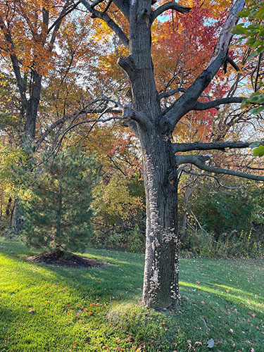
Figure 1. The trunk of this recently deceased maple tree
is covered
with Bjerkandera adusta fruit bodies. Figures
2-6 show photographs of this fungus on the same tree.
Photo © Gary Emberger.
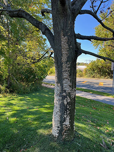
Figure 2. The tree in Figure 1 but from a slightly different
angle.
Hundreds
of fruit bodies developed within a year of
the tree's death.
Photo © Gary Emberger.
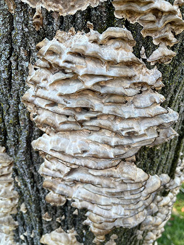
Figure 3. Overlapping caps Bjerkandera adusta. There is
also considerable
lateral fusion of the caps.
Photo © Gary Emberger.
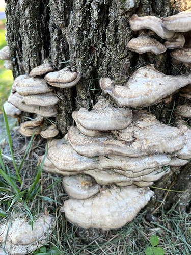
Figure 4.
The upper surface is azonate to faintly zonate.
Photo © Gary Emberger.
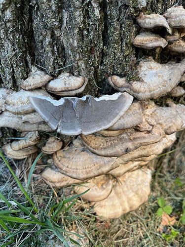
Figure 5. The upper surface contrasted with the grayish
pore surface.
Photo © Gary Emberger.
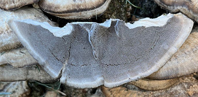
Figure 6. The cracks in the pore surface are artifacts of
tearing off rather than cutting the specimen from the bark.
A pale-colored flesh underlies the gray-colored pore layer.
Photo © Gary Emberger.

Figure 7. Note the dark bruising on the gray pore surfaces.
Photo © Dianna Smith.

Figure 8. The upper surface of this young specimen is
covered by whitish hairs. The cap surface becomes
glabrous with age.
Photo © Gary Emberger.